![]()
![]()
![]()
Lower motor neurons arise in the central nervous system but exit into the peripheral nervous system to innervate muscle, organs and glands. The prototype is the anterior horn cell (less commonly called "anterior root cells"), also known as the final common motor pathway. All cells in the motor cranial nerve nuclei are also lower motor neurons.
Upper motor neurons remain within the central nervous system. All descending moptor fibers that can influence the activity of the lower motor neuron constitute the upper motor neurons. This definition includes both corticospinal (pyramidal) and extrapyramidal fibers.
Damage to upper motor neurons produces:
1. spasticity or hypertonicity (begins after a short period of initial flaccidity)
2. hyperreflexia (begins after a short perior of initial hypo- or arreflexia)
3. extensor plantar responses and other corticospinal signs
4. little or no atrophy, and then only late from disuse
5. no fasciculations
6. loss of strength is incomplete and diffuse
7. loss of superficial reflexes
Damage to lower motor neurons produces:
1. flaccidity
2. hyporreflexia or arreflexia
3. no corticospinal signs
4. quick atrophy (2-3 weeks)
5. fasciculations
6. loss of strength may be severe and in muscles innervated by single nerve/root
![]()
A ganglion is a localized aggregation of neuronal cell bodies in the peripheral nervous system. A nucleus is a localized aggregation of neuronal cell bodies in the central nervous system. The function and termination of all cells within a nucleus or ganglion is the same.
![]()
A tract or fasciculus is a bundle of axons in the central nervous system. A nerve is a bundle of axons in the peripheral nervous system.
A projection is a bundle of central fibers on its way to a target. One tract may have more than one projection.
Spinal roots are bundles of axons leaving the spinal cord. The anterior root carries information out of the cord (motor); the dorsal root carries information (sensory) into the cord. All fibers in the anterior roots are lower motor neurons. A root cell, anterior or posterior, arises from the anterior or posterior horn respectively. Their axons travel in the spinal roots. They are not part of the central nervous system. A column cell has its cell body and axon confined to the central nervous sytem, to which they properly belong. Column cells may be central, internuncial, commissural or associative.
![]()
On cross section, the spinal cord has several divisions. These sections are not artificially separated.
On gross examination, there is a butterfly-shaped central gray area, made primarily of neuronal cell bodies. This gray area is divided into the Rexed's laminae. Each lamina contains a rather homogeneous type of neuron with similar size, function and afferent/efferent relationships to the peripheral and central nervous system. The "wings" of the butterfly extend posteriorly as the dorsal horns and anteriorly as the anterior horns.
The central gray area is surrounded by white matter. Although there are some neuronal bodies in the white matter, it is mainly made of ascending and descending axons. White matter is divided into three funiculi (anterior, posterior and lateral). Funiculi are separated from each other by septae and sulci. The axons may travel ipsilaterally all the time, or cross over to the contralateral side (i.e. may decussate) on their way up or down the neuraxis. These decussations are frequently called commisures.
Different tracts arising in various laminae, and the axons of which travel in various funniculi, decussate at different levels, either in the spinal cord or brainstem.

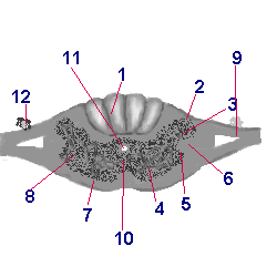
Figure 1. Spinal Cord, transverse section.
1- posterior funniculus (entire area) 2- dorsal root entry zone (DREZ) 3- dorsal horn (left) 4- anterior horn (left) 5- intermediolateral horn (only thoracic and upper lumbar levels) 6- lateral funiculus 7- anterior funiculus 8- anterior horn (right) 9-dorsal root 10- anterior gray commissure 11- central canal 12- dorsal root ganglion.
![]()
There are 5 spinal cord levels:
1. cervical- 8 segments
The cervical spinal segment contains the largest number of fibers in the white matter, making the white matter area the largest (relatively) of the cord. the posterior funiculus is occupied by the posterior columns. The reticular nucleus is seen throughout the cervical region, next to large dorsal horns.
2. thoracic- 12 segments
The T1 to T6 levels contain the posterior columns; at more caudal levels, only the fasciculus gracilis is found.
Only in the thoracic region do we find the lateral horn, containing the intermediolateral column, the source of preganglionic fibers of the sympathetic nervous system.
The dorsal nucleus of Clarke is present throughout the thoracic cord, at the base of the dorsal horn, but is most prominent between T10 and T12.
3. lumbar- 5 segments
The fasciculus gracilis and the dorsal nucleus of Clarke are present. The lateral horn is also present. All three are seen at theh L1-2 levels. The lumbar cord contains the least amount of white matter, both in absolute and relative terms.
4. sacral- 5 segments
5. coccygeal- 1-2 segments
Thus, the spinal cord has 5 levels and, typically, 31 segments. This myotomal segmentation is deterimined by embryologic development, as the limbs migrate and the trunk elongates and turns.
![]()
EMAIL ME AT: SYSTOLE
![]()